Cardiac MRI vs Echocardiography: Key Differences and When to Use Each
Every year, over 15 million echocardiograms are done in the US, but only about 1.2 million cardiac MRIs. Why the big difference? It’s not about which test is better-it’s about knowing when each one shines. Both cardiac MRI and echocardiography are vital for heart health, but they serve different purposes. Let’s break down what each does, when they’re used, and why doctors might choose one over the other.
Cardiac MRIA non-invasive imaging technique that uses magnetic fields and radio waves to create detailed images of the heart without radiation.
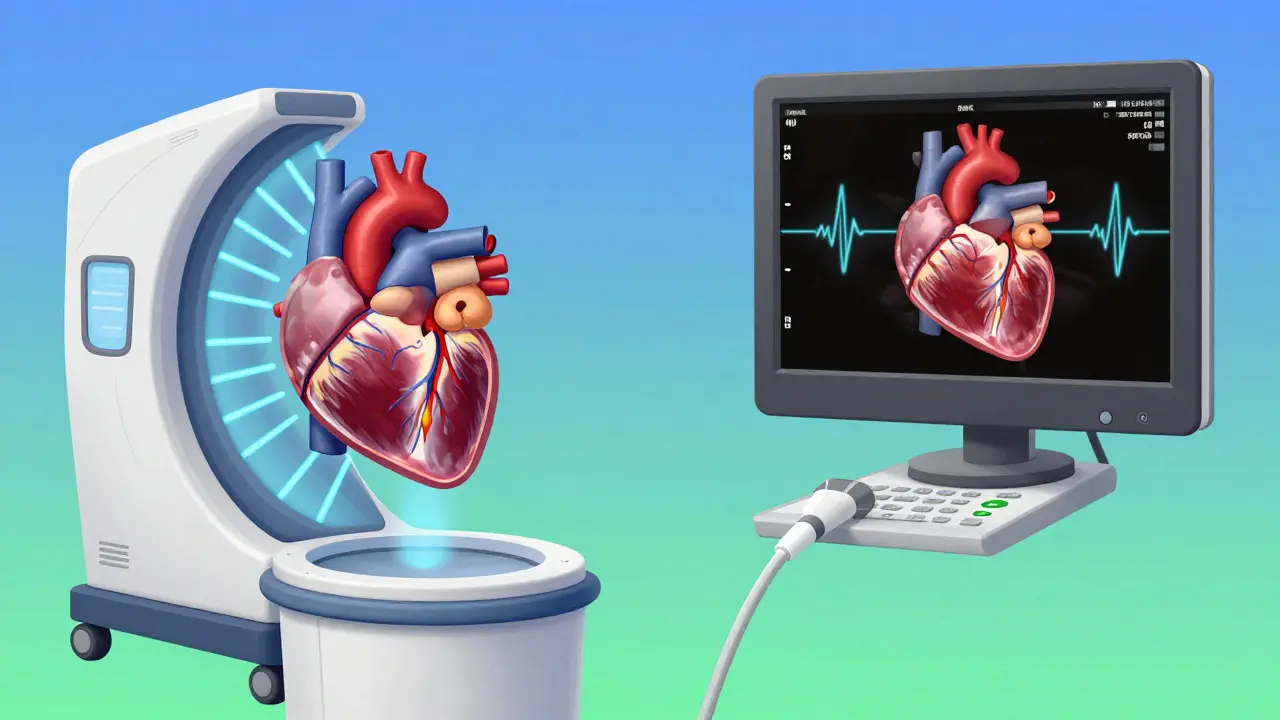
Cardiac MRI, also called CMR, uses powerful magnets and radio waves to take detailed pictures of your heart. It doesn’t use radiation like X-rays or CT scans. Instead, it creates 3D images by measuring how your heart’s tissues respond to magnetic fields. This gives doctors a clear view of your heart’s structure, blood flow, and even the health of your heart muscle tissue. It typically uses magnetic fields between 1.5 and 3.0 Tesla. This high strength allows for very precise measurements. For example, cardiac MRI can measure heart chamber volumes with less than 2% variability between different doctors-much better than echocardiography.
EchocardiographyA common ultrasound-based test that uses sound waves to create real-time images of the heart.
Echocardiography, often called an "echo," uses sound waves to make moving pictures of your heart. Think of it like an ultrasound for your chest. A technician moves a handheld device over your chest, sending sound waves that bounce off your heart. These echoes create real-time images on a screen. It’s quick, painless, and doesn’t involve radiation. Most hospitals and clinics have echo machines, making it the go-to test for initial heart checks. Standard measurements include left ventricular end-diastolic dimension (LVEDD) of 37-56 mm and left ventricular ejection fraction (LVEF) of 50-75%.
| Feature | Cardiac MRI | Echocardiography |
|---|---|---|
| Technology | Magnetic fields and radio waves | Ultrasound waves |
| Accuracy for volume measurements | High (no geometric assumptions) | Lower (requires assumptions) |
| Cost per test | $1,500-$3,500 | $500-$1,500 |
| Accessibility | Requires specialized centers | Widely available |
| Best for | Detail tissue analysis, precise volume measurements | Real-time assessment, initial screenings |
When should you get one test over the other? If your doctor suspects a heart valve problem or needs to check blood flow, they’ll likely start with an echocardiogram. It’s perfect for real-time assessment. But if they need to see if there’s scarring in your heart muscle or measure exact volumes, cardiac MRI is the gold standard. A 2021 study found that echocardiography often underestimates the heart’s pumping ability compared to MRI-by up to 15% in some cases. That difference matters when tracking heart failure treatment.
Real-world examples show why both tests matter. A cardiologist in Boston shared a case where a patient’s echo showed normal heart function, but the MRI revealed hidden scarring. That finding led to early treatment for a condition that could have led to sudden cardiac arrest. On the flip side, in emergency situations like chest pain, echocardiography is invaluable because it’s available immediately at the bedside. As one echo tech put it on Reddit, "I’ve seen countless cases where poor acoustic windows led to inaccurate measurements that were corrected by MRI."
Recent tech advances are making both tests even better. Philips’ latest ultrasound machines use AI to automate measurements, reducing errors. Meanwhile, Siemens’ new 0.55T MRI machines are safer for patients with metal implants, like pacemakers. Experts predict these tools will work together more often in the future-using echo for quick checks and MRI for deeper analysis when needed.
For patients with heart conditions, understanding these tests can help you ask the right questions. If you’ve had an echo but your doctor still isn’t sure what’s going on, they might recommend a cardiac MRI. It’s not about one test being "better"-it’s about using the right tool for the job.
What’s the main difference between cardiac MRI and echocardiography?
Cardiac MRI uses magnetic fields and radio waves to create highly detailed 3D images of the heart, while echocardiography uses sound waves for real-time, moving images. MRI excels at measuring heart volume and detecting tissue issues like scarring, while echo is better for quick, portable assessments of heart valves and blood flow.
Is cardiac MRI safe for everyone?
Most people can safely have a cardiac MRI, but it’s not recommended for those with certain metal implants (like older pacemakers) or severe kidney problems. Newer MRI machines, like Siemens’ 0.55T model, are safer for patients with implants. Always tell your doctor about any medical devices before the test.
How long does each test take?
An echocardiogram usually takes 30-45 minutes and can be done in a doctor’s office. A cardiac MRI typically takes 45-90 minutes and requires a specialized MRI machine. Both are painless, but MRI requires lying still inside a tube-like machine, which can be uncomfortable for some.
Which test is better for detecting heart disease?
It depends on the condition. For valve problems or congenital heart defects, echocardiography is often first. For detailed tissue analysis (like scarring from a past heart attack or myocarditis), cardiac MRI is superior. A 2023 study found MRI detected early heart muscle changes in 90% of cases where echo missed them.
Can I have both tests?
Yes! Many doctors use them together. For example, an echo might show a problem, and an MRI gives more detail. In complex cases like hypertrophic cardiomyopathy, using both tests provides a complete picture. This combined approach is becoming standard in top cardiac centers.
about author

Ashley Hutchins
Cardiac MRI is way better than echo echo is unreliable and misses so much doctors who only use echo are cutting corners Ive seen patients suffer because of it MRI is the only way to get accurate data trust me I know what I'm talking about
Lakisha Sarbah
I get what ur saying but echo is still super useful for many cases MRI isnt always available and can be hard to schedule its about using the right tool for the situation
Ariel Edmisten
Heart echo is quick and cheap for initial checks. MRI gives detailed images when needed. Both have their place in patient care.
Niel Amstrong Stein
I've been thinking about how medical imaging technologies like cardiac MRI and echocardiography reflect our broader approach to healthcare. It's fascinating how we prioritize cost and accessibility over precision in many cases. MRI offers unparalleled detail but requires specialized equipment and time, while echo is quick and portable but has limitations. This tension between thoroughness and practicality is something we see in many areas of medicine. For instance, in cancer screening, mammograms are common but sometimes miss early signs, while MRIs are more accurate but expensive. The same trade-offs apply here. Echo is perfect for initial assessments, especially in emergencies where every second counts. MRI comes in when more detailed analysis is needed, like checking for myocardial scarring or precise chamber measurements. I've read studies showing echo underestimates LVEF by up to 15% compared to MRI, which could lead to incorrect treatment decisions. This is especially critical for patients with heart failure where accurate measurements guide therapy. On the other hand, echo's real-time imaging is invaluable for assessing valve function during procedures. The key is using them together when necessary. Some top cardiac centers already do this, using echo for quick checks and MRI for deeper analysis. Technological advances like AI in ultrasound and lower-field MRI machines are making both tests more accessible. These innovations could bridge the gap between cost and accuracy. Ultimately, the goal is to provide the best care for each patient, not to pit one test against the other. It's about choosing the right tool for the job, which requires understanding both the strengths and limitations of each. We need more education for doctors and patients about when each test is appropriate. This way, we can avoid unnecessary procedures and ensure accurate diagnoses. In the end, it's not about which test is better, but how we integrate them into comprehensive patient care. 🌌
Paula Sa
I really appreciate how you explained the balance between the two tests. It's true that they complement each other. For example, in hypertrophic cardiomyopathy, using both gives the full picture.
Mary Carroll Allen
Echo is great for emergencies but MRI is where it's at for serious stuff. I've seen too many cases where echo missed the mark and MRI saved the day. It's a game-changer for accurate diagnosis.
Joey Gianvincenzi
Your assertion regarding the superiority of MRI in specific scenarios is not entirely accurate. Echocardiography remains the cornerstone of cardiac imaging due to its accessibility and real-time capabilities. Dismissing its utility is unprofessional.
Amit Jain
MRI is overhyped. Echo is just as good for most cases. MRI costs way more and isn't necessary. Doctors just want to bill more. It's all about money.
Sarah B
Echo is good enough for Americans MRI is for wusses
Eric Knobelspiesse
Echo is fine for basic stuff but MRI shows the truth. However, both are limited by human error. We need better tech. But hey, what do I know I'm just a toxic analyst.
Heather Burrows
I don't know why we even bother with both tests. It's all just a waste of time and money. Just do MRI every time.
Ritu Singh
Echo for quick checks MRI for detail. Simple.